Heat That Speaks: how infrared thermography became a quiet ally in sports injury prevention
Infrared thermography, once associated mostly with industrial inspection and engineering, has earned a stable place in sports medicine over the past decade. In professional football clubs across Europe, South America and other major leagues, the technology is now part of daily athlete monitoring, especially during periods of intense training or congested match schedules.
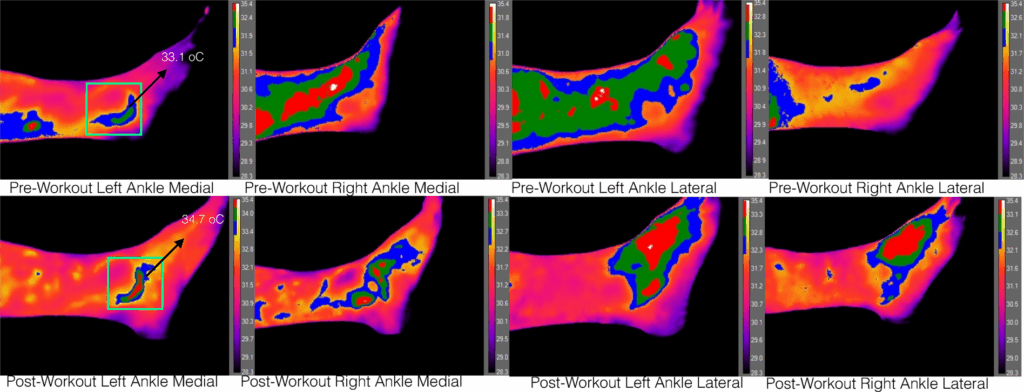
The principle behind the method is straightforward: every area of the body emits heat. When there is irritation, early inflammation, muscular overload or altered blood flow, that region may show a different thermal pattern compared with its opposite side or with the rest of the body. Thermal cameras capture these variations in skin surface temperature – without physical contact and without ionizing radiation – translating subtle physiological changes into visual maps of heat.
In elite football, the goal is prevention. By detecting thermal asymmetries that appear before any noticeable pain, medical teams can adjust training load, increase recovery strategies, direct physiotherapy, or simply keep an eye on the athlete for a few days. Many teams include thermography in their morning screening routine, alongside wellness questionnaires, sleep assessments and light physical checks. The objective is not to diagnose injuries, but to identify early signs that something may be heading in that direction.
For everyday individuals – runners, gym-goers, people undergoing rehabilitation or workers exposed to repetitive strain – the value is similar. Thermography can reveal areas under excessive load, asymmetries after heavy training, delayed recovery patterns or regions that deserve attention before symptoms emerge. Because the exam is quick, non-invasive and free of significant contraindications, physiotherapists, sports physicians and occupational health professionals increasingly use it as a complementary tool alongside clinical evaluation.
The technology itself is accessible to explain: the camera detects natural infrared radiation emitted by the body and converts it into an image. What we see is not the inside of the muscle, but changes in skin temperature that reflect underlying physiological processes such as increased blood flow, muscular tension or early inflammatory response. This is why interpretation must be done by professionals familiar with both the technique and the individual’s context – including training load, clinical history, environmental conditions and recovery status.
Thermography does not replace traditional imaging – such as ultrasound or MRI – and it does not diagnose injuries on its own. Its strength lies in revealing early, subtle changes, prompting simple interventions before bigger issues develop. In elite sport, this can help reduce the incidence of muscle injuries across a season. For the general population, it can mean recognizing overload patterns early enough to avoid prolonged discomfort or time away from training.
As monitoring technologies become more widespread, thermography continues to establish itself as a useful intersection of science, prevention and performance – both inside the stadium and far beyond it.
Ioannou, S. Functional Infrared Thermal Imaging: A Contemporary Tool in Soft Tissue Screening. Sci Rep 10, 9303 (2020). https://doi.org/10.1038/s41598-020-66397-9